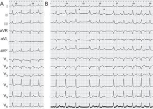
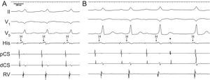
A 28-year-old patient with no structural heart disease was referred for electrophysiological study and ablation to treat an electrocardiographically-documented pattern of ventricular preexcitation. The baseline electrocardiogram (Figure 1A) showed sinus rhythm with a short P-R interval, positive delta wave at leads I, II, III, and aVF, and transition at lead V3, which indicated an anteroseptal or midseptal pathway. Three catheters were positioned: for the coronary sinus (CS) (proximal [pCS] and distal [dCS]), His bundle, and right ventricle (RV). Coinciding with the initial acceleration of sinus rhythm, episodes of second-degree (Wenckebach) atrioventricular block were observed (Figure 1B; asterisks mark blocked P waves), with the peculiarity that ventricular preexcitation remained constant in all the ventricular complexes. This occurred even in the escape complexes that followed the blocked P waves, coinciding with the following sinus P wave. These spontaneous phenomena were diagnostic for the presence of a fasciculoventricular pathway. These are uncommon accessory pathways that connect the bundle of His or its branches with the ventricular septal myocardium. The diagnosis of accessory fasciculoventricular pathway usually requires intravenous adenosine administration or atrial pacing maneuvers. The baseline intracardiac recordings confirmed the existence of ventricular preexcitation, with an HV interval of 10ms (Figure 2A). Thereafter, preexcitation remained the same during progressive prolongation of the P-R interval, and was also observed in the escape beats following the blocked P waves (Figure 2B). Once these accessory pathways have been identified, they do not need to be eliminated because they are not associated with sustained tachycardia.
ISSN: 1885-5857
Impact factor 2023
7.2

