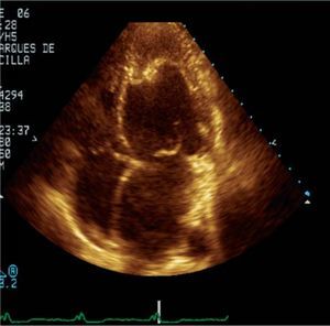
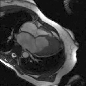
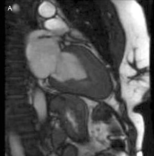
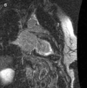
A 54-year-old woman with a diagnosis of apical hypertrophic cardiomyopathy was referred to the cardiac transplant unit for clinical assessment. Her cardiovascular history included hypertension and 2 prior hospitalizations for heart failure. Her anamnesis was notable for an episode of hepatic failure and hypereosinophilia following ingestion of watercress 15 years before. Later, the imaging studies, particularly contrast-enhanced transthoracic echocardiography (Figure 1), disclosed mid-apical obliteration of the left ventricle, which was also observed in dynamic magnetic resonance angiography (MRA) 4-chamber (Figure 2) and 2-chamber (Figure 3A) cardiac views. In the MRA examination, following late passage of gadolinium contrast material, the delayed sequences showed subendocardial enhancement consistent with endomyocardial fibrosis (Figure 3B), which confirmed the diagnosis of Loeffler disease. Following initiation of conventional treatment, the patient remained stable from the cardiovascular standpoint and was managed with close outpatient follow-up and serial imaging studies (transthoracic echocardiography and MRA).

Figure 1.

Figure 2.

Figure 3A.

Figure 3B.