Transesophageal echocardiography has become of increasing importance in guiding structural interventions as more patients with structural heart disease undergo this treatment modality. Depending on the type of intervention, fluoroscopic imaging is not always the most suitable imaging technique to obtain sufficient anatomical information. Thus, echocardiography plays a very important role in intraprocedural monitoring during percutaneous coronary intervention, especially in the treatment of perivalvular leaks, MitraClip device implantation, and left atrial appendage closure.
The EchoNavigator system (Philips) is a recently introduced innovative tool, which automatically fuses X-ray fluoroscopic and echocardiographic images in real time. The movement of the X-ray arc is synchronized with the transesophageal probe of the echocardiogram and the different echocardiographic projections are sliced according to the position of the X-ray source.
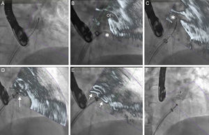
In the Figure A, the X-ray image shows the guidewire and guide catheter in the left atrium. The fused image (Figure B) shows the location of the guide tip (indicated with an asterisk) and its relationship with the left atrial appendage (OI). The Figure C shows the correct introduction of the guide into the atrial appendage, and the deployment of the device (Figure D and E). Finally, the deployment of the device is shown in the fluoroscopic image without fusion (Figure F). This new tool allows different types of images to be fused, thereby helping to facilitate interventions and improve procedural safety.
