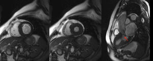
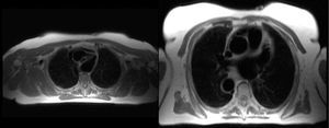
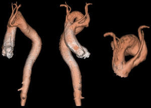
A 69-year-old woman with syncopes and recently diagnosed hypertrophic cardiomyopathy was referred to our imaging unit to complete a cardiac magnetic resonance study. This confirmed the hypertrophic cardiomyopathy (mediobasal septum 17 mm) with left ventricular outflow tract obstruction and anterior systolic motion of the mitral valve (Figure 1). In the anatomical axial planes, we observed an image compatible with double aortic arch (Figure 2), which was confirmed by aortography with contrast medium and posterior 3-dimensional (3D) reconstruction. The 3D images (Figure 3) showed a complete double aortic arch originating in the right subclavian and carotid arteries in the right arch and in the left subclavian and carotid arteries in the left arch. The patient denied symptoms related to this finding (dysphagia, stridor, or dyspnea).


Double aortic arch represents about 1% to 2% of all congenital heart and aortic disorders. It is a complete vascular ring, characterized by a connection between the left and right aortic arches, encompassing the trachea and the esophagus. This condition is usually diagnosed in childhood, and surgical correction has a good prognosis. There have been sporadic reports in adulthood, for which the most common presenting complaints are dysphagia and asthma-like symptoms. Cases of elderly and asymptomatic patients, such as the one reported here, are exceptional.