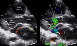
We illustrate the case of a 2-year-old girl presenting with failure to thrive, dyspnea, and systolic murmur. Echocardiography established the diagnosis of cor triatriatum sinistrum with moderate obstruction (arrow) between the upper and lower parts of the divided left atrium with a mean Doppler gradient of 5mmHg (Figure 1; red line = membrane, yellow line = interatrial septum).

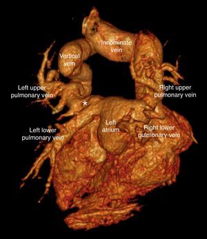
A partial anomalous pulmonary venous drainage of the left upper pulmonary vein to the innominate vein via a dilated vertical vein was also observed. Due to unclear pulmonary venous drainage of all pulmonary veins, the patient underwent coronary computed tomography angiography examination, which confirmed cor triatriatum sinistrum and pulmonary venous drainage of the left upper pulmonary vein to the dilated innominate vein.
A surprising finding was the presence of a connection (*) between the anomalous drained left upper pulmonary vein and the left atrium (Figure 2). The patient underwent surgical correction (membrane excision and ligation of the vertical vein).

Cor triatriatum sinistrum is an uncommon congenital cardiac anomaly accounting for 0.1% of all congenital cardiac defects, in which the left atrium is divided by an abnormal septum. The anomaly may be associated with other congenital cardiac lesions in up to 50% of affected individuals, such as tetralogy of Fallot, ventricular septal defects, and pulmonary anomalous venous drainage.
Most patients are asymptomatic and the clinical manifestation are proportional to the degree of obstruction to pulmonary flow through the membrane, depending on the size of the fenestration.