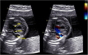
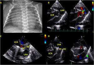
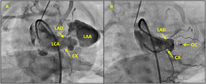

A left coronary artery fistula was diagnosed prenatally at 22 weeks of gestation. An enlarged main left coronary artery (LCA) was connected to the left atrial appendage (LAA) at the site of branching to the left anterior descending (LAD) and circumflex (CX) arteries (figure 1; AO, aortic root). The newborn (birthweight 2510g) had a loud murmur, sinus tachycardia, and significant cardiomegaly (figure 2A). Transthoracic echocardiography confirmed the above-described anatomy (figure 2B). Closure of the fistula was indicated after 15 days due to persisting clinical signs of a significant shunt. Informed consent was obtained from the parents. Retrograde access through the femoral artery was obtained, and angiography of the aortic root was performed (figure 3A, video 1 of the supplementary data). A 4-Fr Amplatzer TorqVue LP catheter (Abbott, USA) was inserted into the LAA using the coronary wire as a rail, and an Amplatzer Piccolo Occluder 9-PDAP-04-02-L (Abbott, USA) was delivered through the catheter. The distal retention disc was kept in the LAA while the central and proximal discs were opened in the LCA. The position of the occluder and patency of the LAD and CX were confirmed by a 10T-D Micro Multiplane Transesophageal Probe (GE Ultrasound, USA) (figure 2C) and by angiography (figure 3B, OC, occluder; video 2 of the supplementary data). Complete occlusion of the fistula and stable position of the occluder were confirmed on the following day by echocardiography (figure 2D). The Piccolo Occluder was originally designed for occlusion of patent arterial duct in premature newborns weighing more than 700 g; therefore, it could be used off-label for the occlusion of a coronary fistula in a newborn.
FUNDING


Work supported by the Ministry of Health, grant ID: 00064203, Czech Republic; conceptual development of research organization, Motol University Hospital, Prague, Czech Republic. There are no other financial relations or relationships with industry to disclose.
AUTHORS’ CONTRIBUTIONSO. Materna: first author, interventional catheterization procedure. J. Kovanda: transthoracic and transoesophageal echocardiography, article revision. V. Tomek: fetal echocardiography, article revision.
CONFLICTS OF INTERESTNone.
Supplementary data associated with this article can be found in the online version, at https://doi.org/10.1016/j.rec.2022.02.007