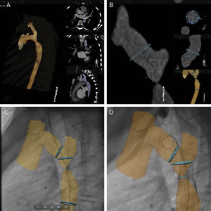
A 15-year-old boy with native coarctation of the aorta was referred for percutaneous stent implantation. Preprocedural arterial-phase computed tomography (CT) datasets were uploaded in a dedicated workstation and subsequently visualized using a novel software package designed for planning and guidance of vascular procedures (VesselNavigator, Philips, Best, the Netherlands) to achieve real-time image fusion between CT datasets and intraprocedural fluoroscopy, facilitating stent deployment (Video of the supplementary material).
One-click segmentation of the aorta was performed to select the anatomic regions of interest (Figure A). Ring markers were placed into the distal transverse arch, stenosis, and the descending aorta to perform standard vessel measurements (Figure B). In addition, “landing zone” reference points were placed as a rough estimate of desired stent length. CT datasets and 2-dimensional fluoroscopic images in 2 perpendicular projections (anteroposterior and straight-lateral) were then coregistered. Manual image alignment was done using bony structures such as the spine and the ribcage, as well as the aortic shadow as reference. Finally, the aortic 3-dimensional-roadmap was used for live guidance (Figure C). Uneventful delivery of a bare-metal stent was performed without residual pressure gradient. Follow-up aortography confirmed a nicely positioned stent (Figure D).

Image guidance using pre-existing CT datasets with traditional fluoroscopy to provide real-time overlay of targeted anatomic structures represents a novel and promising tool in vascular and cardiac interventions. It allows the operator to have a dynamic 3-dimensional roadmap serve as a useful landmark, particularly when dealing with complex vascular anatomies. Furthermore, this imaging modality could shorten the duration of interventions and also reduce contrast and radiation exposure compared with traditional 2-dimensional angiography.