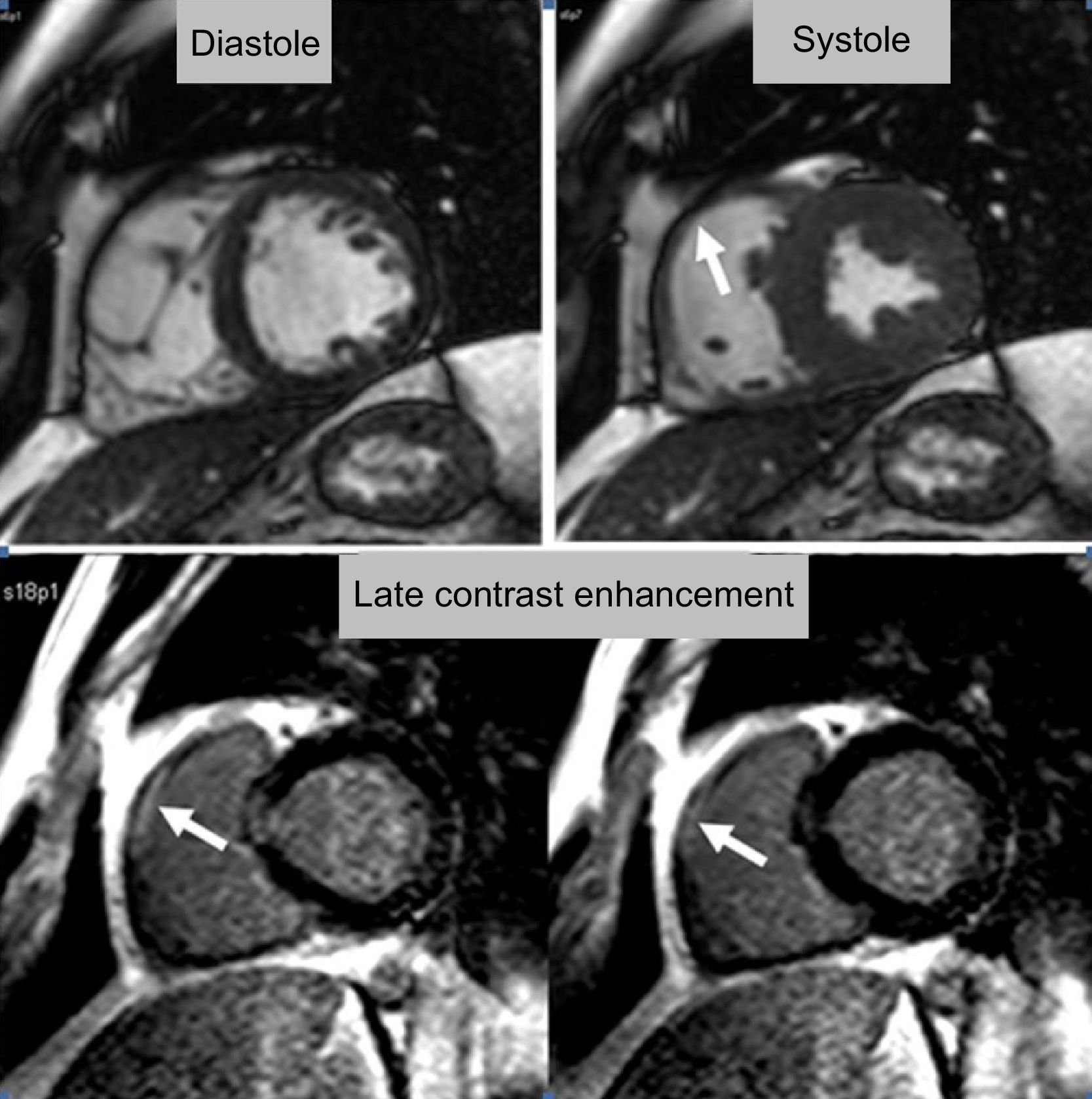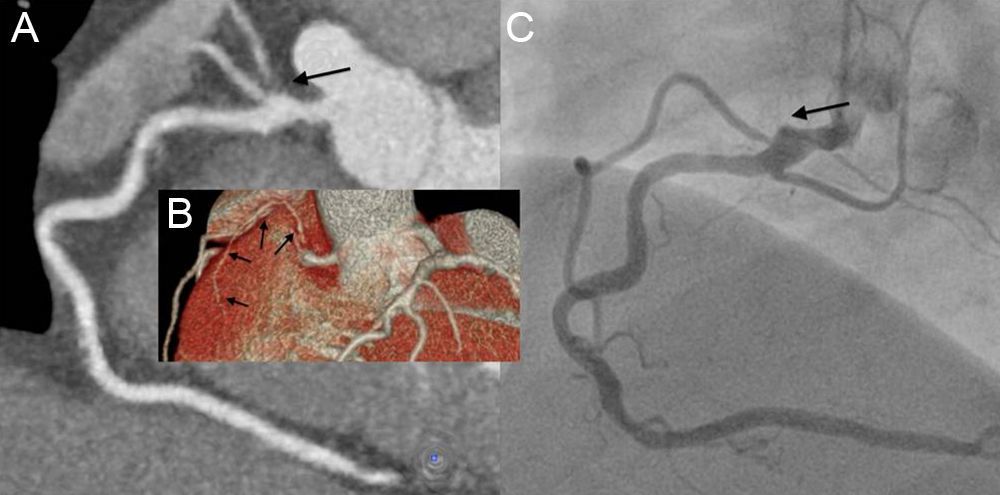

The patient was a 46-year-old man with a history of hypercholesterolemia and obesity, who was admitted to the hospital emergency department with a diagnosis of acute coronary syndrome with ST-segment elevation in leads V1 and V2. He underwent emergency invasive coronary angiography, which revealed a 30% diameter lesion at both the origin of anterior descending artery and the origin of posterior descending artery. Given these findings, the decision was made to complete the study with cardiac magnetic resonance, which showed reduced segmental contractility at the level of the right ventricular outflow tract (Figure 1, arrow in upper panel), coinciding with late contrast enhancement compatible with necrosis (Figure 1, arrows in lower panel). A subsequent noninvasive multidetector computed tomography coronary angiographic study confirmed the presence of the lesions observed during the catheterization and also showed a conus branch in the infarcted region, with thrombotic occlusion at its origin (Figures 2A and B, arrows). This lesion had gone unnoticed in the catheterization, but when the images were reanalyzed, the stump corresponding to the origin of the occluded conus branch could be seen in one of them (Figure 2C, arrow). This case illustrates the high sensitivity of cardiac magnetic resonance for the detection of the regions of necrosis at any site, as well as the utility of combining it with multidetector computed tomography, for an integrated analysis of coronary artery disease.
ISSN: 1885-5857
Impact factor 2023
7.2