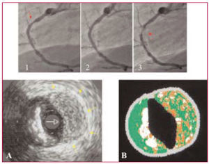
A 64-year-old man underwent coronary angiography for exertional angina with severely positive ischemia tests. The study showed a severe, calcified, focal lesion in the right coronary artery (Figure 1-1). An attempt was made to assess the lesion with intravascular ultrasound (IVUS), but the catheter could not cross it; however, the procedure did reveal a superficial calcification on the plaque, with an angular extension of around 300°.

Figure 1.
With the use of a 3-mm balloon inflated to 18 atm, the lesion was dilated (Figure 1-2); nonetheless, progressive occlusion of the vessel was seen just distal to the original lesion in the confirmatory angiography immediately after the procedure (Figure 1-3). Intravascular ultrasound was performed and at the same location an image was seen consistent with a mural hematoma facing a calcified plaque (Figure 1A). The image provided by radiofrequency analysis (Virtual Histology, Volcano Inc.) showed a diffusely speckled "necrotic core" pattern over the entire thickness (Figure 1B). Although it is not validated in this context, this technique may be able to specifically characterize the fresh blood collection of typical to a mural hematoma.