A 34-year-old man with ST-segment elevation myocardial infarction was referred to our department for primary angioplasty. A coronary angiogram revealed thrombotic occlusion of the ostial left anterior descending artery, with protrusion of abundant thrombotic material into the left main coronary artery.
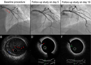
After ineffective thromboaspiration, dilatation with a 2.0mm×15mm balloon was performed and blood flow was restored, although a large thrombus remained (Figure A, red arrows). Intravascular ultrasound demonstrated the presence of an insignificant fissured plaque (Figure B, blue arrows), with abundant attached thrombotic material (Figure B, red arrows). We considered implanting a drug-eluting stent that would reach the left main coronary artery but, given the absence of significant stenosis, the presence of normal flow in an asymptomatic patient, and the risk of distal embolization, this option was ruled out and we decided to administer abciximab and admit the patient to the coronary care unit.

After 5 days of antiplatelet and anticoagulation therapy, coronary angiography was repeated and optical coherence tomography was performed. Both studies revealed partial disappearance of the thrombus, but persistence of the fissured plaque (Figure D, blue arrows). Coronary angiography and optical coherence tomography performed 19 days later showed nearly complete healing of the plaque (Figure F, green arrow) and the absence of thrombus. The patient was discharged from the hospital and, 2 months later, he had experienced no new episodes.
The Figures demonstrate the importance of intravascular imaging in decision making and illustrate the process of vascular healing following rupture of an atherosclerotic plaque. In this case, intravascular imaging avoided the unnecessary implantation of a stent into the left main coronary artery of a young patient, thus reducing the risk of distal embolization and avoiding long-term stent-related events.