To the Editor:
Arrhythmogenic right ventricular cardiomyopathy or dysplasia (ARVD) is a genetically determined heart muscle disease in which the myocardium is replaced by fibroadipose tissue. It is associated with arrhythmias, heart failure and sudden death.1 Typical symptoms include palpitations, dizziness and syncope. Sudden death is often the first sign, especially in young people, and it coincides with some type of strenuous physical activity; in some series, it accounts for up to 20% of all sudden deaths in young adults.2 With the introduction of automatic implantable defibrillators, many of these deaths can be prevented. However, the prevention of arrhythmic death requires timely diagnosis, which depends on the ability of the primary care physician and the specialist to be aware of and to diagnose this disease.
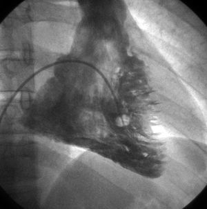
The diagnosis is established on the basis of certain major and minor criteria involving the evaluation of the family history, the depolarization or repolarization-induced electrocardiographic changes, arrhythmias, structural changes and ventricular dysfunction, and on the histopathological features.3 At the present time, there are a number of registries underway for the purpose of analyzing the validity of these criteria and broadening them, if appropriate.4 The typical clinical signs, together with T-wave inversion in leads V1 to V3 present in over 50% of the patients,5 or the appearance of the epsilon wave in 30%,6 raise the suspicion of this disease; however, there are reports of cases in which the electrocardiographic features were less common, but the risk of sudden death was the same.7 We present the case of a 31-year-old man, a professional soccer player, who was referred to our unit because of occasional palpitations. After initial evaluation and the verification of the absence of a family history of heart disease, we examined the electrocardiogram, which revealed negative T waves in leads II, III, and aVF and ventricular premature beats with right bundle branch block (RBBB) morphology (Figure 1). During 24-hour Holter monitoring, several episodes of nonsustained ventricular tachycardia (NSVT) were recorded, as were premature beats with RBBB morphology that appeared to originate in the left ventricular apex. Transthoracic echocardiography revealed a slightly dilated left ventricle with apical akinesia. Given these findings, the patient underwent single photon emission computed tomography (SPECT) to study myocardial work, reaching 20 MET without symptoms, but with frequent premature beats and self-limited episodes of NSVT similar to those recorded during 24-hour Holter monitoring. The scintigraphic images disclosed a fixed apical perfusion defect. In view of these findings, coronary arteriography was carried out, but no lesion of any type was observed; however, ventriculography revealed the presence of left ventricular apical akinesia. Right ventricle was dilated and unstructured, with akinetic/dyskinetic areas and diastolic septal bulge ("stack of coins"; Figure 2). As ARVD was suspected, magnetic resonance imaging was requested. It revealed fatty infiltration of the right ventricle. ARVD with involvement of the left ventricle was diagnosed.
Figure 1. Twelve-lead electrocardiogram recording showing sinus rhythm with T-wave flattening in lead II, negative T waves in leads III and aVF and ventricular premature beats with right bundle branch block morphology.
Figure 2. Right ventriculogram showing akinetic/dyskinetic areas with diastolic septal bulge ("stack of coins") compatible with arrhythmogenic right ventricular dysplasia.
The involvement of the left ventricle in ARVD has been documented previously5; however, electrocardiographic evidence of negative T waves in inferior leads and NSVT with RBBB morphology had not been reported until now. Given that, on many occasions, sudden death is the first sign of this disease, we must be aware of the fact that it may be associated with atypical electrocardiographic features, and it is essential to convey cases like that reported here for the purpose of bringing this cardiomyopathy to the attention of all health care professionals.


