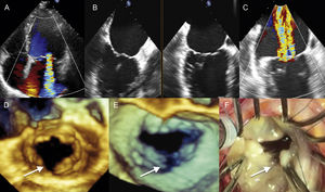
A 71-year-old patient with a history of paroxysmal atrial fibrillation consulted for progressive dyspnea in the setting of a new arrhythmic episode. Transthoracic echocardiogram showed ventricular dysfunction and severe mitral regurgitation, which was considered functional. Electrical cardioversion was performed, achieving normal ventricular function, although severe mitral regurgitation was maintained (Figure A). Transesophageal echocardiogram showed a mitral valve with normal opening, with no evidence of prolapse or restricted leaflets in the orthogonal biplane view (Figure B and video 1 of the supplementary material). Color Doppler confirmed the severity of regurgitation, comprising 2 confluent jets in the bicommissural plane (Figure C and video 2 of the supplementary material). Real-time 3-dimensional images showed a complete cleft (arrows) between the P2 and P3 scallops of the posterior leaflet (Figure D [atrial view] and Figure E [ventricular view], video 3 and video 4 of the material supplementary). No other cardiac abnormalities were observed in the study. The patient underwent surgical intervention, and the echocardiographic findings were confirmed (Figure F).

Although a mitral valve cleft is the most common cause of congenital mitral regurgitation, it is extremely rare in elderly adults, and very few cases of this abnormality have been published. In the majority of patients, myxomatous valvular degeneration is also observed. Three-dimensional echocardiography is useful for diagnosis, and surgical repair is needed if there is significant valvular regurgitation.