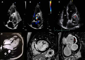
A 40-year-old woman with hypereosinophilic syndrome was referred for cardiovascular screening. A transthoracic echocardiogram demonstrated mildly impaired left ventricle (LV) function and normal right ventricle (RV). The aortic valve was trileaflet with severe aortic regurgitation and a large left sinus of Valsalva aneurysm (SVA) (figure 1A,B, parasternal short-view). An echocardiogram at 1 year revealed a mural thrombus inside the SVA (red asterisk, figure 1C, apical 5-chamber view).

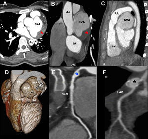
Cardiac computed tomography confirmed a 64 x 55 x 61 mm SVA with a large thrombus, partial compression of left pulmonary veins (LPV), displacement of pulmonary arteries (PA) and coronary arteries (figure 2A,B,C,D) with a left anterior descending artery (LAD) aneurysm (black asterisk) originating in the distal left main artery (LMA) (figure 2E) and right coronary artery (RCA) aneurysm (blue asterisk, figure 2F).

Cardiac magnetic resonance showed focal transmural myocardial infarction in the mid-septum (figure 1E,F). Surgical treatment was decided with performance of aortic root replacement and 4 cardiac artery bypass grafts with left internal mammary artery (LIMA) to the first diagonal branch (D1), Y-graft using saphenous vein graft to distal LAD and first oblique marginal, and saphenous vein graft. Postoperative TTE showed normal aortic prosthetic function, without new wall motion abnormalities.
Three months later, the patient was readmitted for acute coronary syndrome. Cardiac computed tomography showed occlusion of LMA and significant ostial stenosis of LIMA-to-D1. Percutaneous coronary angioplasty to the LIMA anastomosis site was performed. No angina was found during follow-up.
SVA and coronary artery aneurysms are unusual cardiac involvement of hypereosinophilic syndrome. Early treatment should be initiated when cardiac involvement is identified. Surgical revascularization should be carefully planned when there are no significant coronary lesions.
The authors acknowledge the support of the staff in the Echocardiography Department of Hammersmith Hospital (London, United Kingdom)