Dear Editor,
A 67-year-old man had presented 7 months earlier with complaints of flushing, diarrhea and tiredness. Etiologic investigation diagnosed metastatic midgut neuroendocrine carcinoid tumor for which he had trans-arterial embolization and started monthly sandostatin. In follow-up, he developed progressive exertional dyspnea and was referred for carcinoid heart screen.
On physical examination there were systolic and early diastolic right-sided murmurs, increased jugular venous pressure with prominent v wave and pitting peripheral edema.
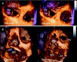
Bidimensional transthoracic echocardiography showed hypomobile fibrotic tricuspid leaflets that resulted in incomplete coaptation and severe regurgitation. The pulmonary valve was not clearly visualized but there was evidence of moderate regurgitation and mild stenosis. Three-dimensional (3D) echocardiography using volume-rendered imaging provided global simultaneous assessment of the three tricuspid leaflets, which were thickened, retracted and immobile, resulting in a fixed orifice (Figure 1A, Video 1). En face volume-rendered imaging of the pulmonary valve, acquired in left parasternal view, also showed thickened hypomobile cusps (Figure 1B, Video 2). The tricuspid subvalvular apparatus and false tendon were also thickened (Video 3). Nevertheless, right ventricular function was normal, as assessed by tricuspid annular plane systolic excursion and right ventricular fractional area change. Speckle tracking strain analysis also showed normal right ventricular systolic deformation.

Figure 1. A: volume-rendered 3-dimensional echocardiography image showing thickened, retracted and immobile tricuspid leaflets in diastole and systole. B: en face volume-rendered image of the pulmonary valve acquired in left parasternal view showing thickened hypomobile cusps in diastole and systole. Ao, aorta; LV, left ventricle; MV, mitral valve; PV, pulmonary valve; TV, tricuspid valve.
Carcinoid syndrome is an uncommon cause of valvular heart disease.1 However, cardiac involvement occurs frequently in patients with this syndrome,2,3 and adversely affects prognosis. This case demonstrates that 3D echocardiography provides comprehensive evaluation of the tricuspid and pulmonary valves in carcinoid heart disease with incremental anatomical detail. Specifically, 3D imaging provides better morphological and functional assessment of right-sided valvular lesions and allows the identification of surrounding structures affected by the pathologic process. Accordingly, this modality may contribute to better recognition of carcinoid heart in clinical practice. Moreover, right ventricular function may be preserved even in the presence of widespread right-sided valvular and subvalvular disease.
Appendix A. Supplementary MaterialSupplementary material associated with this article can be found in the online version available, at doi:10.1016/j.rec.2010.10.009.
Corresponding author: silvamarques.j@gmail.com