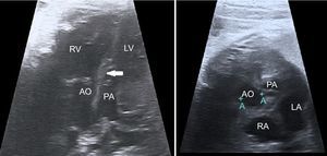
We present an echocardiogram recorded in a 21-week old fetus. The image was indicative of transposition of the great arteries (TGA), with atrioventricular concordance and ventriculoarterial discordance. Additional findings included ventricular shunt (VS) and subpulmonary stenosis. An atypical characteristic was the presence of subpulmonary conus (arrows) and absence of subaortic conus (stars). Only 29 such cases have been published, 22 of which corresponded to postmortem reports. There have been no reports of cases in a living fetus. Subpulmonary conus arises from a mitropulmonary discontinuity (not seen in typical TGA) and the absence of subaortic conus, aortic-tricuspid continuity (Figure 1A, the mark on the image before the selected one is a measurement in 2-dimensional mode; Ao, aorta; LA, left atrium; LV, left ventricle; PA, pulmonary artery; RA, right atrium; RV, right ventricle). The subpulmonary conus appears to be stenotic from its proximal portion. The great vessels are located side by side, with the aorta in a slightly posterior position to the pulmonary artery. The VS is below the subpulmonary conus and closer to the aortic valve.

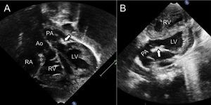
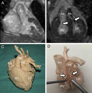
The findings were confirmed by postnatal echocardiography (Figure 2), which ruled out the patient for arterial switch surgery in the neonatal period. Magnetic resonance imaging was performed at 2 months of life to provide more detailed anatomical information and facilitate planning of the surgical strategy (Figure 3A and B). A 3-dimensional model was printed from these imaging data (Figure 3C and D).


To the best of our knowledge, this is the first report in the literature of fetal diagnosis of this uncommon type of TGA and VS. Although the pathophysiological behavior is like any other case of TGA + VS with subpulmonary stenosis, the anatomical characteristics described for this malformation make treatment a challenge. Complementary images and 3-dimensional reconstructions were used to design an appropriate therapeutic strategy.
FUNDINGPart of this investigation was funded by the Instituto de Salud Carlos III, of the Ministry for Science and Innovation, Health Research Project, Health Research Grant no. PI14/00180.