We present the case of a 70-year-old man whose medical history included previous inferolateral myocardial infarction that was not revascularized. Some months prior to his visit, he had undergone transthoracic echocardiography due to exercise angina. This study had shown preserved left ventricular ejection fraction with an undilated left ventricle and absence of valve disease. He had also undergone myocardial perfusion single-photon emission computed tomography, which showed a small area of nontransmural inferoseptal necrosis and minimal inferior ischemia. This was controlled by medical treatment.
The patient attended the clinic for a 3-day history of progressive angina. The electrocardiogram showed ST depression in the inferior and lateral leads, which normalized with nitrate administration. The first ultrasensitive troponin T measurement gave a value of 113 ng/L (< 13 ng/L). Physical examination detected a continuous murmur present in all areas, irradiated from the right lumbar region of the abdomen. This had not been detected previously. On palpation, no pulsatile abdominal mass or thrill could be detected.
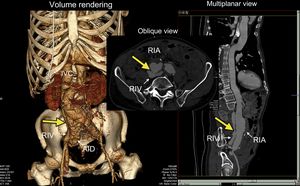
With suspicion of type 2 non–ST-elevation acute myocardial infarction, thoracoabdominal computed tomographic angiography was performed. This study diagnosed a large arteriovenous fistula (Figure, yellow arrow) resulting from spontaneous rupture of an aneurysm in the right iliac artery (RIA) toward the right iliac vein (RIV), with substantial dilatation of the inferior vena cava (IVC).

With this case, we wish to emphasize the importance of careful physical examination for appropriate diagnosis and treatment of patients in cardiology.