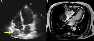
A 75-year-old man who underwent a quadruple coronary artery bypass in 1981 consulted our clinic with dyspnea on moderate exertion. Echocardiography revealed a paracardiac cyst-like mass in the apical 4-chamber view near the right atrium that compressed the lateral atrial wall and had no clear cause (Figure 1A). Cardiac magnetic resonance imaging showed an encapsulated and extracardiac heterogeneous mass with a hyperintense spot corresponding to the patent lumen of a vessel (Figure 1B). Pharmacological stress testing was positive for myocardial ischemia in the right coronary artery territory.

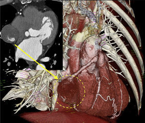
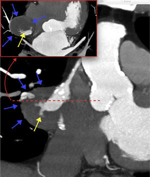
Cardiac computed tomography clarified the origin of this structure, which was the cause of the ischemia. Figure 2 shows a venous graft in the right coronary artery with a saccular aneurysmal dilatation in its middle third (arrow). Figure 3 shows contrast extravasation (yellow arrows) giving rise to the formation of a partially thrombosed pseudoaneurysm cavity (blue arrows).


The incidence of pseudoaneurysms of venous grafts is very low and is higher for grafts placed in the right coronary artery.
Graft dehiscence and pseudoaneurysms can be caused by vascular trauma at implantation, poor surgical technique, and other factors, such as infection and inflammation.
Due to the rarity of this complication, there is no consensus on its optimal treatment, although conservative treatment can be considered in at-risk patients with multiple comorbidities.