An 84-year-old woman with a single-chamber pacemaker and degenerated tricuspid bioprosthesis (Carpentier 26mm, implanted in 2009) was referred to our center for percutaneous valve-in-valve implantation.
An Edwards SAPIEN 3 26mm valve was successfully implanted. Postoperatively, the pacemaker demonstrated normal sensitivity and output parameters and stable impedances. Postprocedural echocardiography showed a pulsatile fluid collection in communication with the right ventricle close to the ventricular pacing lead.
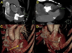
Multislice computed tomography (MSCT) confirmed the presence of a pseudoaneurysm with a dimension of 30 x 14mm in coronal view (Figure 1A) and a neck of 12mm in long-axis view (Figure 1B). Right ventricular systolic pressures measured by echocardiography increased to 55mmHg and MSCT at 9 days showed an increase in pseudoaneurysm size (arrow in Figure 1C) compared with postprocedural MSCT (asterisk in Figure 1D).

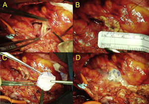
Due to the risk of rupture in the setting of increased right ventricular pressures and pseudoaneurysm size, surgical repair was successfully performed. Intraoperative images of pseudoaneurysm repair show the false aneurysm opened by the scalpel in the right ventricle (Figure 2A), crossing points around the break (diameter 2.5mm) (Figure 2B), double patch (Dacron + bovine pericardium) and “U” crossing points in the patch (Figure 2C) and final view after bonding the patch (Figure 2D).

The likely mechanism for the pseudoaneurysm was increased pacemaker lead tension resulting from fixation of the ventricular lead between prostheses. Prevention of this complication requires careful positioning of the valve, leaving sufficient slack between the tricuspid ring and the point of the right ventricle.