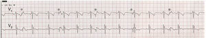
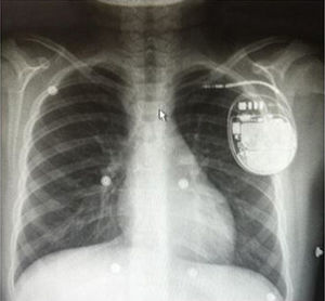
The electrocardiogram showed sinus P waves at a heart rate of about 75 bpm, with a normal PR interval, 1:1 conduction, and QRS morphology indicative of incomplete right bundle branch block. In addition, pacing spikes of the implantable cardioverter defibrillator (ICD) (Figure 1, asterisks) were seen at a heart rate of about 30 bpm (normally the back-up pacing heart rate used in ICD programming for patients not requiring cardiac pacing), compatible with a ventricular sensing failure. These spikes provoked a heterogeneous action potential morphology that had no effect on the atrial and ventricular cycles, indicating capture of an extracardiac structure. Chest radiography (Figure 2) showed dislodgement of the lead toward the pectoral region due to apparent rotation of the device on its sagittal axis, which would produce capture of the chest muscles (correct answer, no. 1).


None of the other options would explain the electrocardiographic findings: in the antitachycardia therapy (option 2), rapid ventricular pacing would be observed; battery depletion (option 3) would not cause any electrocardiographic changes in this situation because the patient had normal sinus rhythm and did not require pacing; lead fracture (option 4) is usually accompanied by noise in the ventricular canal, which would trigger inappropriate discharges due to oversensing.