To the Editor:
We present the case of a pre-term newborn at 33 weeks gestational age with a birth weight of 1730 g, who suffered respiratory difficulty and required non-invasive ventilation without supplementary oxygen and umbilical vein cannulation. On the third day he presented a clinical profile of sepsis with abnormal infectious parameters, and vancomycin and cefotaxime treatment was administered. Evolution was poor, with signs of right heart failure and audible regurgitating systolic murmur 2-3/6 in the tricuspid; the echocardiograph showed a large pediculated vegetation in the right atrium and two small vegetations in the septal tricuspid valve. There was a 5 mm permeable oval foramen with a left to right short circuit. Staphylococcus aureus was isolated in the blood culture.
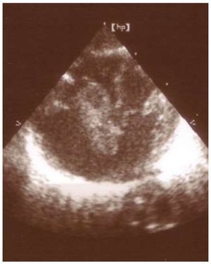
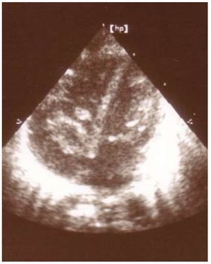
The patient was referred to our centre, where we diagnosed endocarditis of a redundant Eustachian valve with peripheral echo refringencies (Figures 1 and 2) that were compatible with thrombotic vegetation. Antibiotic treatment was maintained during 6 weeks with the addition of low molecular weight heparin, and the patient's evolution was good. The echocardiography before the patient was discharged showed that the vegetation on the Eustachian valve had been somewhat reduced, and that the 2 small vegetations on the tricuspid valve had disappeared.

Figure 1.Four-chamber view in which the large vegetation can be seen protruding from the right ventricle.

Figure 2. Four-chamber view in which the vegetations on the Eustachian and tricuspid valves can be seen.
At 6 months of age, the patient underwent extracorporeal surgery involving total resection through right atriotomy with no post-operatory incidents. Anatomical pathology showed a redundant Eustachian valve with vegetation remnants and thrombosed areas. No microorganisms were isolated in the culture.
The Eustachian valve is an embryological remnant which serves in the foetus to direct the blood flow toward the left atrium through the oval foramen; it can be identified in the ostium of the inferior vena cava and seen on the inside of the right atrium.1 Its presence is infrequent in adults, but echocardiography identifies it in all infants younger than 1 month and in 2/3 of older children; only occasionally is it large and mobile.2
In the literature review, we found a total of 17 cases of infectious Eustachian valve endocarditis, all of which were adult patients. Intravenous drug addiction is the main risk factor for Eustachian valve endocarditis.3 We have found no other recorded case during the neonatal period. The fundamental source of infection in these patients is central catheters, added to the state of physiological immunodeficiency presented by newborns, which is heightened in the case of premature birth.
Of the 17 adult cases we reviewed, 11 required study with transoesophageal echocardiography to make the correct diagnosis.4,5 Both techniques offer similar information in paediatric patients, but due to the limitations of transoesophageal echocardiography at this age, and the fact that the acoustic window offers much better quality than in adult patients, transthoracic ultrasound is always the method of choice; the transoesophageal technique is reserved for specific cases.6-8
With respect to the treatment, we should make special mention of the persistence of the vegetation with signs of peripheral thrombosis (requiring antiaggregant and anticoagulant treatment to date) on a redundant Eustachian valve; after a good initial response to medical treatment, it was managed on an outpatient basis. We opted for a surgical procedure at six months fundamentally because the patient showed no initial response, and was a premature newborn with a very low birth weight. This was done considering the risks involved in extracorporeal surgery, the persistence and large size of the vegetation (which according to later echocardiographic images affected the function of the tricuspid valve) and the risk of embolism.