A 73-year-old man who came to the emergency room because of de novo angina was referred for coronary angiography. The procedure was performed through the right radial artery using a 6-French gauge introducer. The patient received 10mg of verapamil and 5000U of heparin via an intra-arterial route.
At withdrawal of the introducer, a tubular structure was observed at the puncture site. It was thought to be endothelium and, consequently, ligature and resection were performed. Doppler ultrasound study demonstrated patent radial artery flow, and the patient was discharged. At 1 month of follow-up, the radial pulse was barely perceptible, but the patient remained asymptomatic.
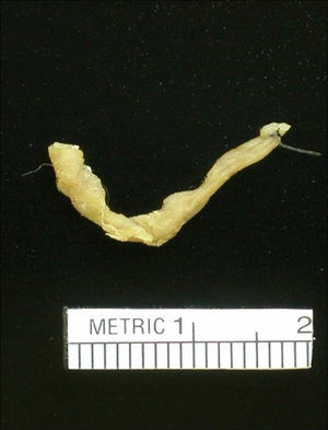
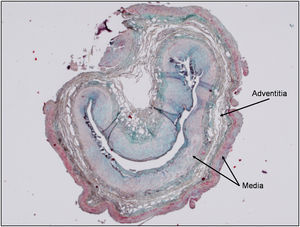
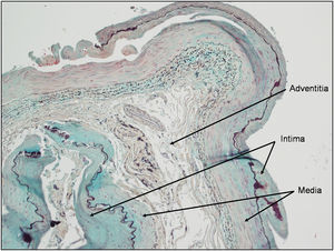
On pathologic study, a 2mm×29mm structure was observed (Figure 1). Microscopy showed that it was a muscular artery. Surprisingly, the three arterial layers were all present. The adventitia was located between the media and the intima, indicating that partial inversion of the artery had occurred (Figure 2, Figure 3).

Figure 1.

Figure 2.

Figure 3.
In the case presented, the radial pulse was present following avulsion of the artery. The reason could be that the arterial fragment was a minor branch emerging from the radial artery or that part of the adventitia had not been extracted, leaving a patent canal.
Total radial artery extraction is an uncommon complication, which, although apparently serious, does not differ from radial artery thrombosis and is rarely accompanied by symptoms of ischemia.