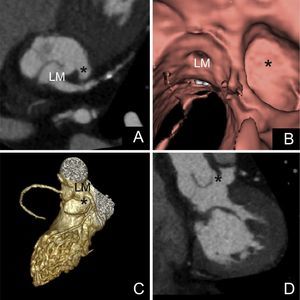
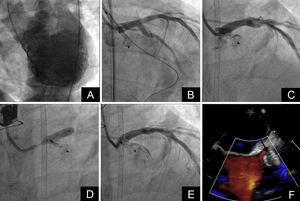
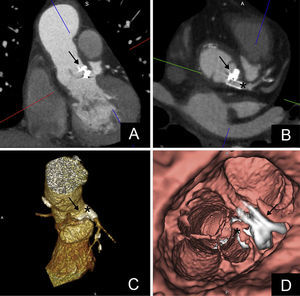
A 59-year old man with a supracoronary graft developed aortic endocarditis, requiring urgent valve replacement through implantation of a biological stentless Sorin aortic valve. Three months after the intervention, he experienced minimal effort angina. A transesophageal echocardiogram showed a periprosthetic leak, causing severe aortic regurgitation. The patient was rejected for a new surgical intervention, and percutaneous closure of the leak was planned. A computed tomography (CT) scan showed a wide perivalvular leak (Figure 1A, Figure 1B, Figure 1C, Figure 1D, asterisk), connected to a periannular cavity situated between the superior left ventricle outflow tract and the aortic wall, close to the left main coronary artery ostium. Ventricular angiography and aortography demonstrated the presence of a pseudoaneurysm formed by dehiscence of the suture line of the prosthetic ring, thus closely related to the leak (Figure 2A, Video 1 of the supplementary material). A wire was advanced to protect the LM, and a 5×10mm vascular plug 2 was placed into the leak (Figure 2A, Figure 2B, Figure 2C, Video 2 of the supplementary material), with protrusion of the proximal disc into the LM (Video 3 of the supplementary material). A 5×15-mm drug-eluting stent was then implanted at the coronary ostium (Figure 2D, Figure 2E, Figure 2F, Video 4, Video 5 of the supplementary material), achieving complete apposition of the plug at the leak entrance, with mild residual aortic regurgitation (Video 6 of the supplementary material). The patient was discharged 2 days after the procedure and remains asymptomatic 6 months later. The follow-up CT scan showed good position of the device (Figure 3, arrow) and the LM stent (asterisk). In complex paravalvular leaks, an exhaustive study of the anatomy is essential to design a tailored strategy for the procedure.
ISSN: 1885-5857
Impact factor 2023
7.2


