A 74-year-old patient with a history of hypertension and chronic renal failure presented at the emergency room with intense chest pain of sudden onset, radiating to the interscapular region. The electrocardiogram showed signs of an enlarged left ventricle with altered repolarization, and plain chest films demonstrated thoracic aorta dilation. Cardiac enzymes were normal and echocardiography showed left ventricular hypertrophy without alterations in regional contractility.
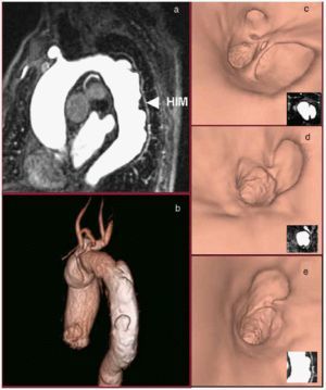
Magnetic resonance (MR) imaging was performed to investigate suspected acute aortic syndrome. The black-blood and bright-blood unenhanced anatomic sequences showed multiple penetrating aortic ulcers producing saccular aneurysms associated with an intramural hematoma (IMH) in the descending thoracic aorta. Gadolinium-enhanced MR angiography with maximum intensity projection (MIP) reconstructions (Figure 1a) and volume rendering (VR) (Figure 1b) with virtual endoscopic vision (Figures 1 c, d, and e) confirmed these findings.

Fig. 1.
The symptoms resolved with antihypertensive drug treatment and there were no new episodes of chest pain. Follow-up magnetic resonance imaging two months after hospital discharge showed a decrease in the size of the hematoma.
Magnetic resonance imaging is a diagnostic tool offering information similar to that provided by multislice computed tomography in patients with acute aortic syndrome in whom the use of iodinated contrast material is contraindicated.