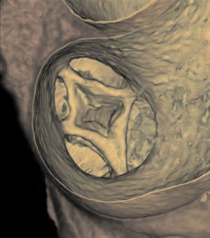
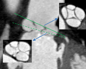
A 63-year-old woman visited our clinic with intermittent chest discomfort and palpitation. Her past medical history included well controlled diabetes and dyslipidemia. Physical examination revealed no abnormal findings. Resting electrocardiography showed normal sinus rhythm. The patient had undergone transthoracic echocardiography examinations several times in the past, which only showed aneurysmal changes of the pulmonary trunk. The right ventricle was of normal size with good systolic function and minimal tricuspid regurgitation. Right ventricular systolic pressure was estimated to be 30mmHg. The patient was further evaluated by electrocardiography-gated 128-slice dual-slice computed tomography (CT). The CT angiography revealed an enlarged main pulmonary artery and a quadricuspid pulmonary valve exhibiting 2 equal and 2 smaller cusps unexpectedly on hollow view seen from above (Fig. 1) and these 4 cusps did not close completely during the mid-diastolic phase on the reformatted images at 74% of the RR interval on the electrocardiogram (Fig. 2). Diagnosis of this rare anomaly is very difficult due to the anatomical disposition of the valve with respect to the thoracic wall and a tendency to be clinically quiescent. The incidence of quadricuspid pulmonary valve was 0.2% among donor hearts in the European Homograft bank. We report a case of a quadricuspid pulmonary valve diagnosed by cardiac CT.


.