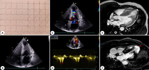

A 49-year-old male was referred to the cardiomyopathy unit due to electrocardiogram changes. He was asymptomatic and denied a family history of cardiomyopathy or sudden cardiac death. An electrocardiogram (figure 1A) showed voltage criteria for left ventricular hypertrophy and diffuse deep T-wave inversion.

Echocardiography (videos 1 and 2 of the supplementary data) revealed severe left ventricular hypertrophy, predominantly involving the mid-segments; hypertrophied, apically-displaced, papillary muscles, causing midventricular narrowing and systolic obliteration (figure 1B); and an aneurysmatic left ventricular apex (figure 1B, white arrowhead). Doppler evaluation revealed dynamic obstruction at the mid-ventricular level (color aliasing – figure 1C); continuous wave Doppler (figure 1D) recorded a pressure gradient of 30mmHg, with a “signal void” pattern, composed by telessystolic (asterisk) and early diastolic (red dot) peaks, which persisted after Valsalva provocation.
Cardiac magnetic resonance showed the apical aneurysm [AA] (figure 1E, white arrowhead), without intracardiac thrombi, and extensive transmural late gadolinium enhancement in the hypertrophied segments and apical cap (figure 1F, red arrowhead). Sarcomeric hypertrophic cardiomyopathy (HCM) was confirmed with a pathogenic variant in the MYBPC3 gene: p.Arg820Gln (c.2459G>A).
Apical aneurysms, an unsolved phenotypic expression in ≤ 5% of HCM patients, are associated with increased mortality. The “Doppler signal void” is depicted: a pattern reflecting abrupt flow cessation across the obliterated ventricle, rendering the recording of the gradient impossible; followed by a paradoxical early diastolic flow, which represents the release of the previously trapped volume. This may explain the adverse effects produced by mid-ventricular obstruction (even in the absence of high recordable velocities on echocardiography), which may be linked to AA development. Since AA has prognostic implications in HCM, recognition of this sign is paramount to pursue multimodality imaging investigation.
The patient consented to this publication.
FUNDINGNone.
AUTHORS’ CONTRIBUTIONSM. Brandão: conception, writing of the manuscript and selection and editing of image material; A. Marchi: selection and editing of image material, revision of the article; I. Olivotto: supervision and manuscript revision.
CONFLICTS OF INTERESTNo conflicts of interest to declare.
Supplementary data associated with this article can be found in the online version, at https://doi.org/10.1016/j.rec.2022.12.009