
A 33-year-old woman presented an anterior myocardial infarction treated with fibrinolytics. On coronary angiography performed a few days later, a dissection of the left anterior descending artery (LAD) was observed, with no lesions in other vessels. The patient was stable and showed no symptoms; hence revascularization treatment was not performed at that time. One week later the patient returned to the hospital with angina and transient ST segment re-elevation from V2 to V6. A new coronary angiography was undertaken. The results were similar to those seen previously, except for an image of a double lumen with an intimal flap from the proximal segment of the LAD to the distal portion, with contrast retention and grade II TIMI flow (Figure 1).

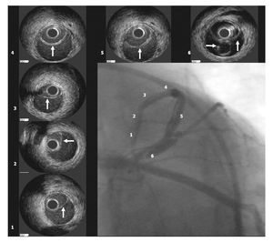
Figure 1
Coronary stent implantation was chosen to treat the dissection; nevertheless, during the procedure it was difficult to determine whether the guidewire was advancing through the true or false lumen. With the assistance of intracoronary ultrasound, it was possible to locate the position of each segment and observe the dissected intima, indicated by the arrows in Figure 1. Two overlapping drug-coated stents, measuring 3×32 mm (distal) and 3.5×2 mm (proximal) were implanted (Figure 2).

Figure 2
In the case presented, intracoronary ultrasound allowed us to: a) confirm the diagnosis, b) determine the location of the guidewire in the true lumen, c) determine the length and diameter of the vessel segment treated, and d) confirm the outcome following stent implantation.