A 60-year-old man with a history of smoking, hypertension, and an earlier episode of prolonged angina, presented to our hospital with progressive dyspnea. He showed signs of heart failure in the physical examination and an electrocardiogram showed sequelae in the anterior leads.
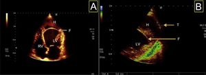
An echocardiogram was performed, showing akinesia in the left anterior descending territory, with a mobile linear intracardiac image consistent with flap dissection (Figure 1A; F, flap; H, hematoma; LV, left ventricle; RV, right ventricle) with involvement of the medial segments of the anterior septum, inferior septum, anterior wall, and entire cardiac apex, giving rise to an apical neochamber. Images of the interior of this neochamber were consistent with thrombosis (Video 1 of the supplementary material). The findings were interpreted as intramyocardial dissecting hematoma. In the lateroapical segment, a continuity solution of 3mm in diameter was observed (Figure 1B; T, tear) corresponding to a tear in the myocardial wall giving rise to a localized pseudoaneurysm.

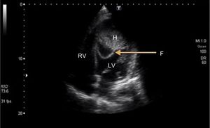
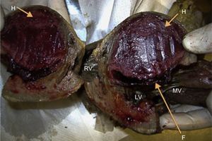
On the sixth day after admission, the patient showed ventricular tachycardia requiring electrical cardioversion; subsequent echocardiography showed progression of the neochamber (Figure 2), with a highly mobile dissection flap (Video 2 of the supplementary material) and the patient subsequently died. The autopsy showed intramyocardial hematoma and dissection flap formed from the endocardium and part of the myocardium (Figure 3; MV, mitral valve, mid-ventricular view).

