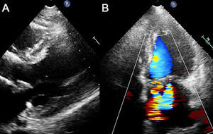
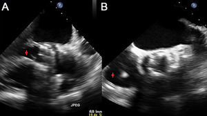
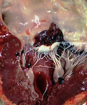
A 71-year-old hypertensive man, who was an exsmoker with respiratory and renal comorbidities, presented with severe, symptomatic aortic stenosis. Due to his high surgical risk (EuroSCORE of 20.5%), we successfully implanted a LOTUS Boston Scientific percutaneous aortic valve transfemorally, with suitable depth. Implantation of a definitive pacemaker was also required, due to complete atrioventricular block. Two months later, the patient was admitted to hospital for acute pulmonary edema, and a new grade V/VI systolic murmur was auscultated in mitral focus. The transthoracic echocardiogram revealed a thickened and perforated anterior mitral leaflet, causing severe mitral regurgitation (Figures 1A and B). Transesophageal echocardiogram showed that the anterior mitral leaflet had heterogeneous echogenicity and irregular margins, suggestive of an abscess (video 1 of the supplementary material). The prosthetic aortic ring and its leaflets were thickened and had a mobile and filiform structure compatible with vegetation (Figure 2A and video 2 of the supplementary material). Moreover, vegetation was visible in the pacemaker lead (Figure 2B and video 3 of the supplementary material). Serial blood cultures isolated Streptococus gallolyticus. In view of the diagnosis of early infective endocarditis of the aortic valve complicated by severe mitral regurgitation due to valve rupture and pacemaker lead endocarditis, we discussed the case with the cardiac surgery team, who ruled out surgery due to the high risk. The patient died 48hours later due to severe sepsis and heart failure. Clinical autopsy confirmed the diagnosis (Figure 3). This is the first known case of infective endocarditis in a patient with a LOTUS percutaneous aortic valve.
ISSN: 1885-5857
Impact factor 2023
7.2


