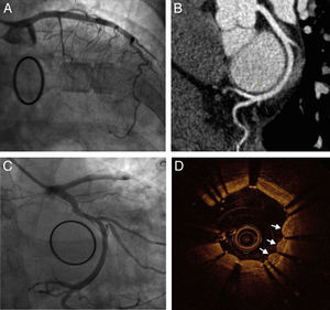
The patient was a 51-year-old woman who was being studied after having a middle cerebral artery stroke. Echocardiography resulted in a diagnosis of mitral valve papillary fibroelastoma, and the decision was made to operate on the valve. Following resection of the fibroelastoma, valve reconstruction, and ring implantation, intraoperative ultrasound revealed the presence of moderate mitral regurgitation, and we decided to replace the mitral valve with a mechanical prosthesis. During the immediate postoperative period, the patient was found to have hemodynamic instability and ST-segment elevation in leads I, III, and aVL. Emergency coronary angiography showed occlusion of the mid circumflex artery (Cx) (Figure A) that had not been observed in the preoperative coronary angiography (Figure B). A guidewire was advanced and, despite considerable difficulty, it was made to cross the region of the lesion with a balloon; high-pressure predilatation was performed with 2-mm and 2.5-mm balloons and adequate expansion was achieved, but there was immediate elastic retraction. Finally, a 3×28-mm drug-eluting stent was implanted, with good angiographic results (Figure C). Optical coherence tomography revealed a peculiar scalloped aspect around the stent struts (Figure D, arrows), which indicated extrinsic compression of the coronary artery and confirmed the diagnosis of Cx occlusion due to compression/traction of the surgical suture.

The Cx runs along the anterolateral commissure, and the coronary sinus is adjacent to the annulus next to the posterior mitral leaflet, both in the atrioventricular groove. Thus, it is possible to damage the Cx during mitral valve annuloplasty or replacement. This has been reported more often with left dominant coronary artery circulation (as in our patient, Figure B) or codominance, as there is less distance between the vessel and the annulus.