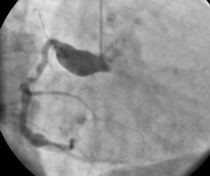
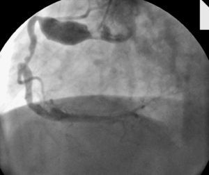
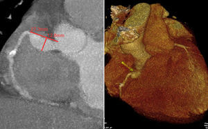
A 64-year-old woman with diabetes consulted for angina at rest. Seven years previously she had experienced anteroapical myocardial infarction. The patient underwent deferred coronary angiography, which showed a significant lesion in the middle third of the right coronary artery (RCA) and an absence of angiographically significant stenosis in the left coronary tree. During the procedure, an image consistent with iatrogenic dissection of the right sinus of Valsalva was observed. Both lesions were managed conservatively. The patient presented repeated episodes of heart failure and progressed to left bundle branch block on electrocardiography and left ventricular dilation with extensive anteroseptal akinesia, an ejection fraction of 21%, and moderate mitral failure on transthoracic echocardiography. Lateral and inferior ischemia was detected on Tc-99m MIBI single photon emission computed tomography, and a new coronary angiography was performed. A huge aneurysm was detected in the ostial-proximal RCA, with likely origin in the right sinus of Valsalva (Figure 1, Figure 2), an absence of significant lesions in the left coronary tree, and a significant calcified lesion in the middle third of the RCA (already described on previous catheterization). Multislice computed tomography confirmed the presence of a huge aneurysm with no proximal neck, a fact that ruled out percutaneous treatment (Figure 3), and severe calcification in the middle third of the RCA. Because percutaneous treatment of the aneurysm was excluded and the patient presented high surgical risk, a decision was made for conservative management.

Figure 1.

Figure 2.

Figure 3.
Coronary aneurysms present in 0.15% to 4.9% of patients undergoing coronary angiography, with the main causes being arteriosclerosis in adults and inflammatory disease in children. Nevertheless, very few cases of “giant” coronary aneurysms (>2 cm diameter) have been described. Interventional coronary procedures are included among the causes of this condition. We describe the case of a giant aneurysm of the ostial-proximal RCA in a patient with a history of sinus of Valsalva dissection, which occurred during diagnostic coronary catheterization.
Corresponding author: sfcoroleu@hotmail.com