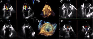
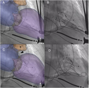
In the implantation of the Tendyne mitral prosthesis for the treatment of severe mitral regurgitation (figure 1A), advanced imaging techniques are essential for success. After placing the delivery sheath in the left atrium (LA) (figure 1B,C), the prosthesis is advanced to the end of the sheath and partially released (figure 2A,B). At this point, it is essential that the prosthesis is aligned in such a way that the upper part of its atrial aspect is oriented toward A2 (figure 1D). To ensure this, en face 3-dimensional echocardiography can be used, and this can be corroborated using fusion imaging with computed tomography. This allows assessment at any time of the position and orientation of the prosthesis, which has a radio-opaque marker (figure 2C, arrow) that must remain oriented toward A1. Fusion imaging allows visualization of the left atrial orifice (figure 2C, asterisk, video 1 of the supplementary data) which indicates the lateral zone of the LA, allowing the desired orientation to be achieved. Once positioned and oriented correctly, the delivery sheath is withdrawn until it makes contact with the annulus and the prosthesis is deployed; this can be confirmed on transesophageal echocardiography and also on fusion imaging. If the position is correct, the prosthesis is anchored to the apex with the dedicated pad and tension is applied to the tether to achieve the desired result (figure 1E,Fvideos 2 and 3 of supplementary data).
CONFLICTS OF INTEREST

R. Estévez-Loureiro is a consultant for Abbott Vascular. The other authors have no conflicts of interest.