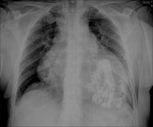
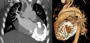
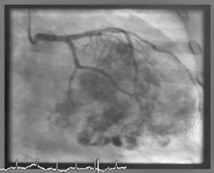
A 66-year old woman with hypertension was referred to our center for heart failure and chest pain. The electrocardiogram showed atrial fibrillation, right bundle branch block and left anterior hemiblock. On chest radiography, the cardiopericardial silhouette was considerably enlarged and marked calcification was seen in the region of the left ventricle (Fig. 1). Transthoracic echocardiography disclosed left ventricular wall thickening with moderate systolic dysfunction, a large intramyocardial calcification that generated reverberation artifacts and limited the quality of the image, moderate mitral and tricuspid regurgitation, and severe pulmonary artery hypertension. On computed tomography, massive myocardial calcification was seen, mainly at the left ventricle and interventricular septum (Fig. 2). Coronary angiography ruled out significant stenosis and showed the severe calcification of the left ventricular myocardium (Fig. 3, videos 1-3). Endomyocardial biopsy confirmed the final diagnosis of calcified endomyocardial fibrosis, based on findings of myofibrillar hypertrophy and endomyocardial fibrosis bands. Surgical resection of the calcified fibrotic tissue was discarded because of the considerable extension of the process. The patient was discharged with medical treatment including loop diuretics, beta-blockers, and angiotensin II receptor agonists. She died some months after by progressive heart and kidney failures.


.