
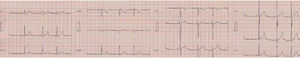
An 11-year-old boy with no history of interest had experienced 4 episodes of palpitations in the previous year and a half. The first 3 of these were short-lived and resolved spontaneously. In the fourth, he attended his local hospital, where the electrocardiogram shown in Figure 1 was recorded. The tachycardia resolved abruptly with vagal maneuvers. Three weeks later, a resting electrocardiogram was recorded (Figure 2). Echocardiography showed mild-moderate tricuspid valve regurgitation, with the regurgitation jet originating from the septal leaflet. The patient was referred to the arrhythmias unit of his referral hospital.


What is the most likely diagnosis?
- 1.
Ventricular tachycardia originating in the right ventricular outflow tract.
- 2.
Sinus tachycardia with frequency-dependent left bundle branch block.
- 3.
Left posterior fascicular ventricular tachycardia.
- 4.
Antidromic supraventricular tachycardia with Mahaim fibers connecting the right atrium and ventricle.
Submit your diagnosis to this ECG Contest at http://www.revespcardiol.org/electroreto/71/3. The diagnosis will follow in the next issue (April 2018). #RetoECG.