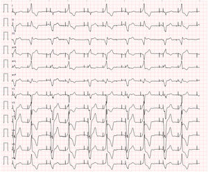
The patient was a 63-year-old man with a history of dilated ischemic heart disease. He had undergone several revascularizations, both surgical (1996) and percutaneous, with interventions for restenoses in the right coronary artery, most recently in 2015. In 2017, the patient underwent ablation because of sustained monomorphic ventricular tachycardia and he received a dual-chamber implantable cardioverter-defibrillator. In the most recent follow-up visit, no arrhythmic episodes were detected. The leads were performing correctly (appropriate atrial and ventricular threshold and impedance, P-wave detection); VP was 100%. In September 2019, he attended the clinic with an echocardiogram showing left ventricular dilatation and an ejection fraction of 37% with inferoseptal hypokinesia and inferoposterior akinesia. The primary complaint was intermittent claudication. The patient reported no dyspnea or exercise-induced angina, orthopnea, paroxysmal nocturnal dyspnea, palpitations, or syncope. The paced electrocardiogram is shown in figure 1.

What was occurring?
- 1.
VDD ventricular pacing alternating with APVP
- 2.
Intermittent conduction via a septal accessory pathway
- 3.
Ventricular extrasystoles with constant coupling
- 4.
Dislodged atrial lead that alternately paces the atrium or the ventricle
Submit your answer to http://www.revespcardiol.org/en/electroreto/73/06. The answer will be published in the next issue (July 2020). #RetoECG.