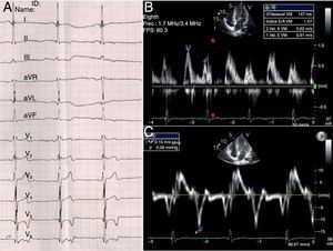
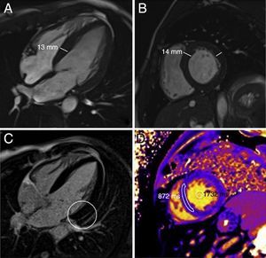
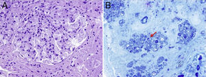
We report the case of a 35-year-old man without systemic hypertension but with a family history of hypertrophic cardiomyopathy who sought medical attention due to malleolar edema. Electrocardiography showed negative T waves in V4-V6 (Figure 1A) and echocardiography showed left ventricular hypertrophy (LVH) but normal diastolic function and Doppler tissue velocity (Figure 1B and 1C). Cardiac magnetic resonance imaging (MRI; Figure 2A and 2B) also showed LVH with preserved left ventricular ejection fraction and without gadolinium retention on late enhancement (Figure 2C). The native T1 relaxation time (Figure 2D) was 872ms, less than normal, with normal extracellular volume. Even in patients with hypertrophy, focal gadolinium retention, or decreased S’ velocity by Doppler tissue imaging, a reduced native T1 can indicate early myocardial involvement in Anderson-Fabry disease (AFD). Given a finding of proteinuria in the nephrotic range and undetectable levels of alpha-galactosidase, renal biopsy was performed, which confirmed AFD by revealing glomeruli with vacuolated hypertrophic podocytes in hematoxylin-eosin staining (Figure 3A) and birefringent particles in the podocytes (arrow with polarized light) (Figure 3B).


In AFD, the alpha-galactosidase deficiency causes multiorgan accumulation of sphingolipids, with cardiac involvement in 90% of patients. Less than 60% of AFD patients have LVH and not all patients show the characteristic gadolinium retention in the lateral basal segment of the left ventricle, as in our patient (Figure 2C, circle). In contrast to amyloidosis and hypertrophic cardiomyopathy, there is a significant reduction in the native T1, with values<940ms effectively identifying 90% of patients; the lower the value, the greater the hypertrophy. Given these findings, the patient's mother was reevaluated and subsequently diagnosed with AFD.