A 10-month-old patient was referred to the Pediatric Cardiology Department to investigate inspiratory laryngeal stridor since birth and difficulty swallowing semi-solid and solid foods since the age of 6 months.
In the physical examination the child showed adequate weight and stature, and inspiratory laryngeal stridor, without dyspnea or cyanosis. The cardiovascular examination, electrocardiogram, and color Doppler echocardiogram were normal.
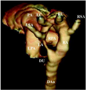
Extrinsic airway and digestive tract compression by a vascular ring was suspected. Esophageal contrast radiography disclosed compression of the upper third of the esophagus (ESO). Helical CT angiography of the heart and great vessels (Figure 1) showed a double aortic arch: one posterior and right-sided (RA), of considerable diameter, from which the right subclavian artery (RSA) and right carotid (RC) artery emerged separately, and along its course, another anterior, hypoplastic left-sided aortic arch (LA) with a large bulge, from which the left carotid (LC) artery and left subclavian artery (LSA) emerged.

Figure 1.
Moreover, a prominent ligamentum arteriosum (DU) inserted at the most distal portion of the anterior arch, before the point where the arch reaches the descending aorta (DAo), completing the vascular ring.
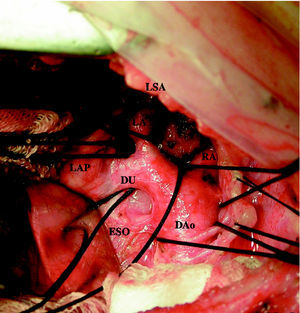
The patient underwent surgical treatment (Figure 2), consisting of resection of the entire portion of the anterior arch from the left subclavian artery to the descending aorta, including the ligamentum arteriosum, thereby freeing the esophagus and trachea.

Figure 2.