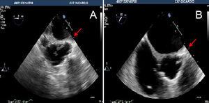
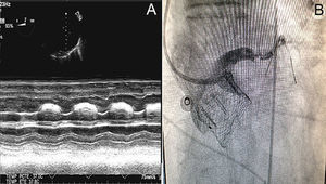
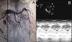
A 74-year-old male with hypertension, type 2 diabetes mellitus, diabetic nephropathy and chronic alcoholic liver disease, presented with severe, symptomatic aortic stenosis. Aortic valve replacement surgery was ruled out due to the high risk and we opted for transcatheter aortic valve implantation (TAVI). A previous coronary angiography showed a severe obstructive lesion in the proximal segment of the left anterior descending artery, which was revascularised with two drug-eluting stents. The patient was admitted to hospital for a scheduled TAVI. During the procedure, while we were implanting a 27 mm LOTUS (Boston Scientific, Natick, Massachusetts, United States) aortic prosthesis transfemorally, the transoesophageal echocardiography showed the appearance of a mobile, thick structure with irregular and broad movements in the ascending aorta, compatible with a thrombus (Figure 1A and video 1A of the supplementary material). This structure was attached to the left main coronary artery (Figure 1B and video 1B of the supplementary material). The patient simultaneously suffered ST-segment elevation on the cardiac monitor and severe hypotension (systolic pressure 50 mmHg), which required inotropic support. Transoesophageal echocardiography showed dilatation of the left ventricle with anteroapical akinesia and severe systolic dysfunction (Figure 2A and video 2A of the supplementary material), therefore we proceeded with an emergency coronary angiography. The left main coronary artery was selectively probed with a guide catheter, revealing thrombotic occlusion of the distal left main coronary artery (Figure 2B and video 2B of the supplementary material). Thrombus aspiration and dilatation were performed with a balloon, achieving a notable improvement in flow. Final flow was TIMI 3 (Figure 3A and video 3A of the supplementary material). In the final transoesophageal echocardiography, the left ventricle was visualised with global systolic and segmental functions preserved (Figure 3B and video 3B of the supplementary material).
ISSN: 1885-5857
Impact factor 2023
7.2


