Spontaneous coronary artery dissection is an uncommon cause of acute myocardial infarction. Coronary artery intramural hematoma is considered within this group of conditions, even though there is no intimal rupture. Angiographic diagnosis of coronary hematoma is complex because the lesion is located within the vessel wall, where contrast does not penetrate.
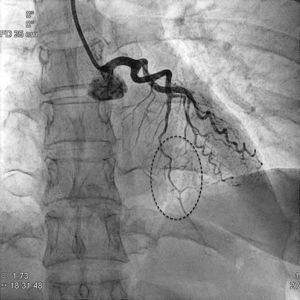
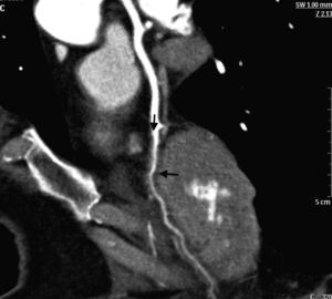
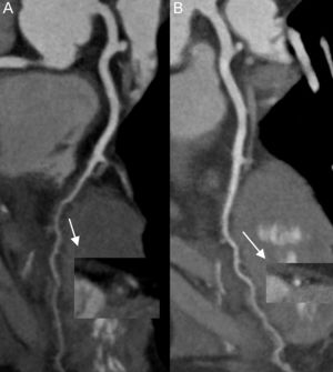
We present the case of a young woman who experienced an ST–segment-elevation acute myocardial infarction of the anterior wall following an episode of stress. Coronary angiography (Figure 1) showed marked narrowing of the left anterior descending artery in its middle third to distal segment (dashed outline), which raised suspicion of intramural hematoma. Because of the noninvasive nature of computed tomography angiography (CTA) relative to intracoronary imaging modalities, multislice CTA was used to confirm the diagnosis and perform follow-up. The CTA image (Figure 2) showed a reduction in the vessel lumen diameter, and additionally, diffuse wall thickening (arrows) with no plaques. In Figure 3, CTA of the hematoma in orthogonal and curved multiplanar images compares the first study, showing diffuse, concentric thickening (Figure 3A, arrow) with the follow-up at 45 days, showing disappearance of the hematoma (Figure 3B, arrow). Repeating CTA was useful to confirm the diagnosis and seems advisable when there are uncertainties. In this case, multislice CTA confirmed the suspected diagnosis and ruled out differential diagnoses such as atherosclerosis and coronary vasospasm.


