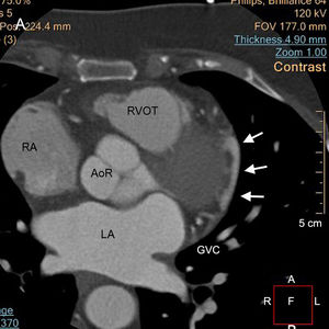
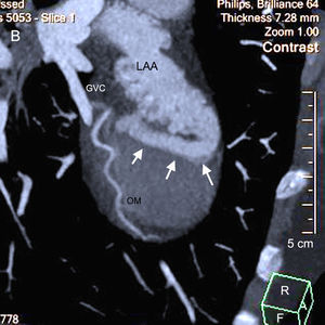
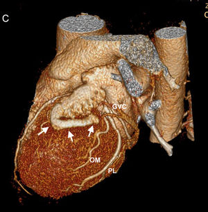
A 72-year-old woman was referred for noninvasive coronary angiography with multidetector computed tomography due to prolonged chest pain at rest and moderate functional limitation due to dyspnea; she had hypertension, obesity, and hyperthyroidism. Conventional cardiac stress tests were invalid due to low levels of exertion but an outpatient rhythm study (Holter) revealed dynamic ST changes. With metoprolol and nitroglycerin premedication, helical CT with retrospective reconstruction was acquired, timed with the electrocardiogram and during the administration of 80mL of nonionic contrast agent, with a dynamic 64-slice system and iterative reconstruction (Brilliance iCT 64; Philips Healthcare; Best, The Netherlands). The acquisition was processed using proprietary software (Philips IntelliSpace) in accordance with the recommended technical standard. In addition to significant atherosclerotic disease of the left anterior descending artery and right coronary artery, the axial images showed an abnormal vascular structure in the upper part of the obtuse cardiac margin (Figure 1, arrows; GVC, great cardiac vein), at the level of the left atria (LAs) and right atria (RAs), the aortic root (AoR), and the right ventricular outflow tract (RVOT). Via maximum pixel intensity multiplanar reconstruction (Figure 2) and volumetric rendering (Figure 3), the structure was concluded to correspond to a fistulous tract (arrows) connecting the distal end of the left atrial appendage (LAA) with the GVC, next to the obtuse marginal (OM) and posterolateral (PL) arteries. The absence of venous or right chamber dilatation indicated a trivial shunt. Noniatrogenic fistulas of the LAA are infrequent, and their influence on interventional occlusion procedures is unclear.
ISSN: 1885-5857
Impact factor 2023
7.2


