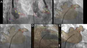
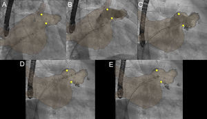
Percutaneous closure of the left atrial appendage is an alternative for patients with atrial fibrillation and a high risk of bleeding or contraindication for anticoagulation. The procedure is performed under transesophageal or intracardiac echocardiographic guidance. The size of the closure device is chosen according to prior measurements or those taken during the 2-dimensional or 3-dimensional echocardiographic procedure or computed tomography. Computed tomography is of particular interest given its noninvasive nature and high spatial resolution. Integration of computed tomography with the fluoroscopy display is possible with fusion systems (HeartNavigator, Philips). After segmentation by chambers and centering using 2 aortographic views (Figure 1A), the movement of the light arc is synchronized and allows both types of image to be superimposed. Figure 1 shows the implantation of an Amulet device (St. Jude Medical) from the start of deployment (Figure 1B), lobe (Figure 1C), relation to circumflex artery (Figure 1D), until the final outcome after release of the device (Figure 1E). Similarly, Figure 2 shows different steps of implantation of a Watchman device (Boston Scientific) with a pig-tail catheter inside the left atrial appendage (Figure 2A), anatomical correlation with 2 angiographic projections (Figure 2B and Figure 2C), start of deployment (Figure 2D), and final outcome prior to release (Figure 2E). Fluoroscopy and computed tomography fusion imaging has been used for percutaneous implantation of aortic valve prostheses and apical approach for closure of leaks or ventricular pseudoaneurysms. To the best of our knowledge, this is the first reported case of its use in percutaneous closure of the left atrial appendage.
ISSN: 1885-5857
Impact factor 2023
7.2

