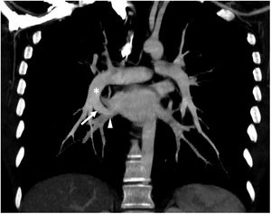
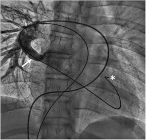
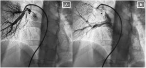

A 16-year-old male with no relevant medical history attended the emergency department with dyspnea, headache, and severe hypoxemia, largely when standing, with baseline saturation of 70% to 75%. He was admitted for study of platypnea-orthodeoxia syndrome. During admission, transthoracic echocardiography was normal; computed tomography (figure 1) revealed an arteriovenous fistula (arrow) that connected the right pulmonary artery (asterisk) with the right inferior pulmonary vein (arrowhead). After this finding, diagnostic catheterization confirmed the existence of this fistula (figure 1, arrow), in addition to a complex anatomy that hindered the advance of the catheter via an angled and stiff Guide Wire M (Terumo Europe, Belgium) (figure 2, asterisk). Catheterization was subsequently performed to close the fistula with a 4-Fr access through the right femoral artery. From this site, a pigtail catheter (Cordis Corporation, United States) was inserted for hemodynamic monitoring. Through a 6-Fr access site in the right femoral artery and with the help of a JR4 catheter (Cordis Corporation) and a guide extension catheter, the sheath was advanced and a 12 × 16-mm vascular Amplatzer Plug II device (Abbott Medical, United States) was implanted in the fistula. The absence of early contrast agent passage from the right inferior pulmonary artery (figure 3A) was verified and delayed filling was evident in the left atrium (figure 3B). After the procedure, saturation was 99% on standing and decubitus, without dyspnea. Follow-up radiography revealed correct placement of the device. At 8 months of follow-up, the patient was asymptomatic and his saturation was within normal limits.


Informed consent was received from the patient for the performance of the complementary tests and procedure and the publication of the clinical case.
FUNDINGNone.
AUTHORS’ CONTRIBUTIONSAll authors contributed to the development, drafting, and revision of this work.
CONFLICTS OF INTERESTWe do not have conflicts of interest.