Full English text available from: www.revespcardiol.org/en
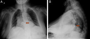
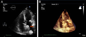
A 93-year-old patient presented to the emergency room of our hospital with fever. Twenty years earlier, she had been incidentally diagnosed with left atrial myxoma measuring 7mm in diameter, with limited mobility attached to the atrial septum. At the time, the patient refused resection surgery and did not return for follow-up. The chest radiograph revealed cardiomegaly and a rounded calcified image on the cardiac silhouette (Figure 1A and B, arrows). Transthoracic echocardiography (Figure 2A and B, arrows) showed a calcified mass measuring 1×5 cm attached to the atrial septum with an associated acoustic shadow of limited mobility, consistent with a calcified left atrial myxoma (Video of the Supplementary Material). She was diagnosed with a urinary infection. In view of the clinical progression, her age, and her prior refusal of surgery, a conservative approach was chosen.


Usually, myxoma is resected after diagnosis. This is clearly the preferred approach when the patient is symptomatic. This case is of interest given the doubts about the approach to take in asymptomatic patients at high surgical risk or those who refuse surgery. Echocardiographic findings such as a smooth surface and calcification suggest lesion stability and limited progression. Calcification is an infrequent finding in atrial myxoma. Visualization of myxoma in a radiograph because of calcification is a rare occurrence.