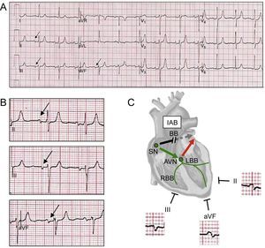
A 71-year-old male patient presented to the emergency department with palpitations, chest discomfort, and rapid atrial fibrillation requiring cardioversion with a 100 J synchronized shock. Electrocardiography performed after cardioversion (Figure A) revealed sinus rhythm with P-wave duration ≥ 120ms and biphasic morphology (+/-) in the inferior leads (Figure B), raising suspicion of Bayés’ syndrome. Bayés’ syndrome is characterized by abnormal electrocardiography findings (advanced interatrial block in sinus rhythm) and an increased risk of paroxysmal supraventricular arrhythmias. The prolonged P-wave duration and the negative final component in the inferior leads, indicated activation of the left atrium in a caudal to cranial direction due to block in the Bachmann region (Figure C; AVN, AV node; BB, Bachmann bundle; IAB, inter-atrial block; LBB, left bundle branch; RBB, right bundle branch; SN, sinus node). Advanced interatrial block has been found to be a predictor of atrial fibrillation recurrence in many clinical scenarios, including postelectrical and pharmacological cardioversion and postpulmonary vein or cavotricuspid isthmus ablation, in patients with advanced heart failure and in Chagas’ disease. The patient was treated with amiodarone and oral anticoagulation and had an unremarkable course at 15 months. Awareness of the electrocardiography pattern in Bayés’ syndrome may permit early treatment, preventing new episodes of tachyarrhythmia and cardioembolic events.
ISSN: 1885-5857
Impact factor 2023
7.2
