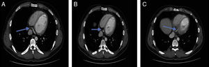
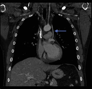
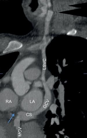
A 28-year-old man with a history of Hodgkin's lymphoma presented with productive cough to the hospital. Contrast enhanced computed tomography (CT) of the thorax was performed to assess for lung pathologies. Incidentally, the CT revealed a lack of connection between the coronary sinus (CS) and right atrium (Figures 1A–C) with a persistent left superior vena cava (LSVC) (Figures 2 and 3; GCV, great cardiac vein; LA, left atrium; MCV, middle cardiac vein; RA, right atrium).


The CS is a clinically important structure given that it serves as an access for various electrophysiological procedures and cardiac surgeries. Because many interventions, including left ventricular or biventricular pacing, mapping procedures, ablations of arrhythmias, targeted drug deliveries and stem cell therapies make use of the CS, it is important to identify anatomic variations and ensure appropriate preprocedural planning.
In very rare cases, atresia of the CS ostium is associated with a persistent LSVC without any connections to the left atrium, as is the case here. In these cases, the LSVC carries blood retrogradely from the coronary system via a bridging vein to the right superior vena cava and eventually drains into the right atrium.
It is important to recognize this anomaly when use or ligation of the LSVC or pacemaker implantation is planned. If this variation is not recognized, ligation can interrupt the CS drainage, causing coronary venous hypertension, myocardial congestion, and even death.