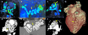
A 36-year-old asymptomatic woman underwent 4D-flow cardiac magnetic resonance (CMR) after an abnormality was observed on electrocardiogram (negative T-waves in V1-V3), to rule out cardiomyopathy (1.5T CMR, optima-450W-GE, 4D-flow; Gadovist gadolinium, 0.15 mmol/kg; matrix, 172 × 172; FOV, 42; VENC, 150 cm/s; bandwidth, 62.50; NEX, 4; voxel size, 2.4 × 2.4 × 2.4mm) (figure 1A-C and video 1 and video 2 of the supplementary data). This showed that the right coronary artery (RCA) had an anomalous origin from the pulmonary artery (ARCAPA), with coronary artery ectasia. Cardiac computed tomography (CT) was then performed to confirm the diagnosis and give a more precise anatomic assessment (RevolutionTM-CT256-GE, USA; contrast, Iopamidol 370 mg/mL) (figure 1D-G). Here we compare the images from the 2 techniques of this rare congenital anomaly.

Cardiac MR showed that the left coronary artery had a physiological origin from the left sinus of Valsalva, with an ectatic and tortuous left anterior descending artery (LAD) (figure 1A, yellow arrow; Ao, aorta). The RCA, also ectatic, had an anomalous origin from the main pulmonary artery (mPA) (figure 1B, posterior descending artery, yellow arrow, and figure 1C, origin of RCA, yellow arrow). The 4D-flow technique (video 1 and video 2 of the supplementary data) demonstrated retrograde flow from the RCA to the mPA (figure 1C, yellow arrow). Cardiac CT (figure 1D-G) confirmed the diagnosis revealing a communication between the posterior descending artery and the LAD (figure 1E, orange arrow).
The 4D-flow (video 1 and video 2 of the supplementary data) was key for a full assessment, and, with single volume acquisition, allowed us to evaluate the origin and trajectory of the coronary arteries and analyze the Qp/Qs (1.1), rule out other associated shunts, visualize the direction of flow at the RCA origin, and perform direct quantification of the shunt.