
We present the case of an 81-year-old woman with severe calcific aortic stenosis (Figure 1A) referred for surgical valve replacement. Immediately after valve replacement (Figure 1B), an aortic annular thickening (asterisk) and an abnormal ovoid echolucent collection at the left atrial (LA) posterior wall (arrowhead) were noted under intraoperative transesophageal echocardiography. Four-dimensional view (Figure 1C) also revealed an unusual trigone configuration (asterisk) with LA wall molding (arrowhead). Midesophageal transesophageal echocardiography multiplane view (Figure 1D) at the end of the procedure was remarkable for the presence of a large LA pseudomass (arrowhead).

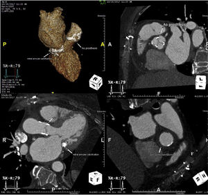
Conservative therapy with protamine administration was decided, with an uneventful recovery and nearly complete resolution at the first week (Figure 2, showing periaortic hematoma [asterisk] at computed tomography).

Extensive periaortic hematoma is a rare complication following aortic valve procedures, either associated with surgery or percutaneous implantation. It is caused by perforation of all 3 aortic layers and is related to annular calcification, protruding plaques of atheroma, prosthesis oversizing, and hypertension during weaning from cardiopulmonary bypass. Early recognition at transesophageal echocardiography is essential for appropriate management through blood pressure control and protamine administration, and this event generally self-limited under serial imaging. Additionally it may allow a differential diagnosis with LA dissection. Despite being more frequently caused by atrioventricular junction injury following mitral valve surgery, this may occur during aortic valve procedures, from inadvertent disruption of LA endocardium. A new chamber with or without communications into the LA, typically appearing as a false echolucent blood filled cavity, supports the diagnosis of LA dissection, a more lethal unforeseen complication.