To the Editor,
In a recent article in Revista Española de Cardiología, Azar et al1 report the results of a study of the volume and function of left atrium (LA) by means of 3-dimensional echocardiography (3D-echo). The study subjects are selected from a noncardiology patient population and volumes and maximum and minimum diameters were tabulated, as were segmental volume changes, thus providing a database that could be of value for comparisons involving populations of cardiology patients. The authors acknowledge in the "Limitations" section that "the 3D-echo analysis of the LA was performed using a computer application designed for the LV [left ventricle]," but, in addition, it should be stressed that the anatomic molds generated by 3D-echo are by no means representative of the structure and anatomic configuration of the LA; thus, the parameters measured are probably very far from representing the real volume and function of this chamber.
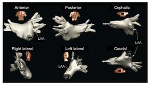
The shape of the LA generated by 3D-echo is ovoid and quite regular; however, the real anatomy is very different (Figure). The LA has a maximum cross-sectional diameter (side-to-side) that goes completely undetected in 3D-echo examinations and other ultrasound techniques. The shape of the LA appears more like 2 truncal-conical volumes of differing "height," with the vertices in the vestibules of the pulmonary veins and the bases merged toward the mitral valve. The limit of these vestibules with respect to the veins is imprecise. The ovoid appearance of the chamber is in no way representative of reality.

Figure 1. Electroanatomic mapping (Navx®) of left atrium (LA) and the pulmonary veins in a patient with paroxysmal atrial fibrillation with no structural heart disease. Note the highly irregular shape of LA, with a large side-to-side diameter (left-to-right) that is not visualized in any of the echocardiographic projections. LAA indicates left atrial appendage; MV, mitral valve.
Magnetic resonance studies show that the side-to-side diameter can increase in size, together with the diameter of the superior pulmonary veins, while other atrial dimensions do not change.2 On the other hand, as would be expected, a good correlation has not been found between the LA diameters measured by echocardiography and the volume of the chamber measured by computed tomography.3
The significance of these data concerning the volumes and function of LA obtained by means of 3D-echo should be interpreted with caution and not without comparing them first with those obtained with imaging techniques capable of defining the complete anatomy of the LA throughout all the phases of its cycle. Meanwhile, magnetic resonance and computed tomography appear to be the only imaging techniques that can enable us to advance with a steady gait at the present time in the understanding of the mechanisms of atrial disease in patients defined until now as "free of heart disease" and "free of atrial dilation," especially in the definition of the anatomic and pathophysiological bases of problems with a marked clinical impact, such as atrial fibrillation.
Dr Francisco García-Cosío has received remunerations for training activities from St. Jude Medical.