The past decades have seen an increase in infections of cardiac implantable electronic devices (pacemakers, implantable cardioverter defibrillators, and cardiac resynchronization therapy devices), and the number of infections appears set to increase further. Prominent factors underlying this trend are the growing number of indications for device implantation and the older age of individuals carrying these devices, with the associated burden of accompanying diseases, mainly kidney failure. Physicians who prescribe and fit these devices are duty bound to familiarize themselves with the prevention, diagnosis, and treatment of these infections.
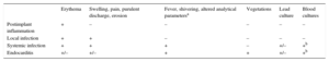
Cardiac device infection can be local (pocket infection) or systemic, and device implantation can also provoke local inflammation; distinguishing among these conditions can be challenging (Table). Postimplant inflammation manifests as erythema near the incision site within 30 days of implantation. This inflammation is caused by mechanical injury and can be difficult to distinguish from local infection. Local infection is indicated by signs of infection in the pulse generator pocket, including swelling, pain, purulent discharge, fistula formation, and skin erosion with or without device extrusion. Device extrusion is always a sign of infection. Systemic infection is further accompanied by systemic symptoms such as fever, shivering, a blood count typical of bacterial infection, and sometimes a positive blood culture. Systemic infection accompanied by vegetations in the device leads or any of the right valves indicates device endocarditis, which is also increasing, both in absolute patient numbers1 and as a percentage of the total number of right endocarditis cases.2
Clinical Conditions Affecting Cardiac Implantable Devices and the Pulse Generator Pocket
Once a diagnosis is confirmed as local infection, systemic infection, or endocarditis, it is crucial to act fast; the entire apparatus, including the pulse generator and leads, must be removed as soon as possible. The most frequent infecting microorganisms are coagulase-negative staphylococci, which are typical in infections of foreign material. These microorganisms are highly adherent, frequently form biofilms that make them resistant to antibiotics, and easily infect the device leads from the pulse generator. Exclusive antibiotic therapy without lead extraction is therefore ineffective against most infections.
It can be very difficult to distinguish between local infection and postimplant inflammation. Preliminary data indicate that these clinical situations can be distinguished by a combination of positron emission tomography and computed tomography.3 If validated, this approach would help in selecting the appropriate therapy in each case: lead extraction for pocket infection and symptomatic treatment for postimplant inflammation. It is nevertheless important to bear in mind that positron emission tomography does not distinguish between inflammation and infection, and findings should therefore be interpreted in the context of the patient's clinical presentation.
It is essential to carefully evaluate the need for device replacement. In some studies, up to a third of patients did not require a new device.4 When a new device is needed, it should be implanted on the opposite side of the chest to the original device after new blood cultures confirm the elimination of any initially blood-culture-positive infection.
Leads should be extracted percutaneously, even when large vegetations are present. This process can lead to complications, and should therefore be undertaken by experienced practitioners.5 In centers with a high patient volume, there is a very high probability of a successful, complication-free outcome. Surgical extraction is associated with more complications, related not only to the procedure itself but also to the patient's age and comorbidities. In the early years of treating cardiac device infection, surgery was recommended when large vegetations were present, but a large vegetation is no longer considered a contraindication for percutaneous extraction. Bacterial vegetations released to the right circulation can cause pulmonary embolisms, but these tend to be asymptomatic and are less aggressive than surgery.
It is important to adhere to recommended timings for the antibiotic treatment, which will obviously be guided by an antibiogram when blood cultures are positive. Antibiotic treatment should be maintained for 10 to 14 days for local infection, 14 days for systemic infection, and 4 to 6 weeks for endocarditis. When blood cultures are negative, the antimicrobials used should be active against the most frequent causes of cardiac device infection: coagulase-negative staphylococci and Staphylococcus aureus.
Several of these observations are reinforced by the results of an excellent study published by Gutiérrez Carretero et al. in Revista Española de Cardiología.6 The study presents experience with 325 cardiac device infections treated by a multidisciplinary team of cardiac surgeons, cardiologists, infectious disease specialists, and anesthesiologists. The number of patients treated and the low reported mortality (1% for local infection and 8% for systemic infection) are testament to the team's expertise in this condition. However, these figures may have been influenced by the low number of patients at high risk of failed extraction or complications: only 8.3% of patients were fitted with cardiac resynchronization therapy devices. The most frequently used device removal method was percutaneous traction, leading the authors to affirm the effectiveness of this extraction technique. Some of the key strengths of the study are the high number of patients included, the rigorous protocol—regarding both the definition of infections and the antimicrobial treatment, and the follow-up of 95.7% of patients for at least 1 year (median, 36 months). Several of the study's findings confirm the conclusions outlined above: a) percutaneous traction is the treatment of choice for both local and systemic infections; b) the presence of vegetations does not contraindicate percutaneous extraction (of the patients with vegetations, only 9% [5 out of 53] had embolic complications, none of which had clinical repercussions or showed a relation to vegetation size); and c) it is difficult to distinguish between local and systemic infection; in this series, positive lead cultures were obtained from half of the patients with ostensibly local infection.
The study also raises several controversial issues related to the treatment of cardiac implantable device infections. For example, the authors’ distinction between acute, delayed, and late infections seems somewhat arbitrary and, moreover, lacks clinical and therapeutic interest.7 The time elapsed since device implantation is not associated with any substantial differences in the causative microorganisms that would justify differential antimicrobial treatment in the absence of positive cultures.
Needle aspiration of the generator pocket is not an accepted diagnostic method, and an earlier analysis cautioned against this procedure.8 Reaching an etiological diagnosis is of course important for selecting the appropriate antibiotic treatment. However, it is unclear what benefit aspiration provides in this context. If there are signs of pocket infection, the pulse generator should be removed and samples taken from the surrounding tissue and the leads. If the pocket infection diagnosis is uncertain, aspiration can itself cause infection.
Another discussion point is that all the devices were removed in the cardiac operating theater under general anesthesia and with orotracheal intubation. In experienced centers, however, cardiologists can carry out device removal in the catheterization laboratory, with no need for general anesthesia or orotracheal intubation.9 Over several years at our center, this approach has yielded results very similar to those reported by Gutiérrez Carretero et al.6 These authors also reported a very high rate of device replacement; only 4% of patients did not receive a replacement device, compared with as many as 33% in other series.4 This high replacement rate may have been influenced by the 1-stage device removal and replacement procedure used in many patients. In our view, careful evaluation of the need for device replacement is essential in the management of cardiac device infection, and frequently shows replacement to be unnecessary.
This 1-stage procedure for device removal and contralateral replacement is the most controversial and novel aspect of the study by Gutiérrez Carretero et al. study. The 1-stage procedure was performed in 72% of patients undergoing device replacement, with very few of them experiencing reinfection (Gutiérrez Carretero et al., Table 8).6 In patients with systemic infection, the reinfection rate with this strategy was in fact lower than when the procedure was carried out in 2 stages. Despite contrary expert opinion expressed in several guidelines, the authors provide convincing data supporting this strategy. The major limitation is the need to postpone device removal until blood cultures are negative (“typically at 1 week” according to the authors). Therefore, in the study, 89% of patients with systemic infection will have experienced a minimum 1-week delay before device removal, as this is the percentage of study patients with initially positive blood cultures (Gutiérrez Carretero et al., Table 3).6 We believe that an infected device should be removed as soon as possible and that decisions should be individualized, taking account of factors such as the level of dependency on the device for maintaining a normal heart rhythm, the duration of antibiotic therapy, and the patient's clinical status. Several groups have reported increased survival when the device is removed in the days immediately following diagnosis.10,11 This unresolved issue warrants further investigation in a randomized study.
In summary, this study highlights the importance of cardiac implantable device infections in day to day practice and emphasizes the need for complete device removal (including the pulse generator and leads) and the benefits of doing this via the percutaneous route. We take note in the interest of our patients.
CONFLICTS OF INTERESTNone declared.