Dear sir:
The study of the causes of stroke points increasingly to the heart,1 especially in young patients. Among these causes are rheumatic valve disease and dilated cardiomyopathy.
The purpose of this Letter to the Editor is to report the case of a 15-year-old girl who presented acute ischemic stroke. The examination revealed arrhythmic heart sounds but no evidence of murmur. At that point in the evaluation, atrial fibrillation was detected and chest radiography demonstrated an increased cardiothoracic ratio that suggested dilated cardiomyopathy.
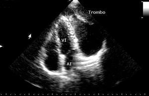
In view of this suspicion, she was referred to our echocardiography laboratory. At the beginning of the study, we observed an echolucent image posterior to left ventricle (LV), as well as mild mitral stenosis. Continuing with the echocardiographic frames from the apical four-chamber view (Figure), the echolucent image seen previously extended laterally to LV. Using color-coded and pulsed Doppler echocardiography, we observed a septal defect between LV and left atrium (LA). Another finding that helped to confirm the diagnosis was the presence of spontaneous echo contrast and thrombi in the chamber.

Figure. Four-chamber apical view showing left ventricle (VI) and left atrium (AI) in relation to the atrial appendage aneurysm, which contains thrombi.
With this diagnosis, the patient was referred to a cardiac surgery center, where surgical treatment for giant aneurysm of left atrial appendage was successful.
To the surprise of students beginning medical training, LA is not situated on the left, but in the middle of the chest. It is the most posterior chamber of the heart.1 Most patients with LA aneurysm present arrhythmia or abnormal silhouette on chest radiograph.2,3 Its diagnosis by means of fetal echocardiography has been described in the literature.4
Correspondence: Dr. L. Torres.
Servicio de Microbiología. Hospital Miguel Servet.
Paseo Isabel la Católica 1, 3. 50009 Zaragoza. España.